Cells, Free Full-Text
Por um escritor misterioso
Descrição
High-resolution 3D images of organelles are of paramount importance in cellular biology. Although light microscopy and transmission electron microscopy (TEM) have provided the standard for imaging cellular structures, they cannot provide 3D images. However, recent technological advances such as serial block-face scanning electron microscopy (SBF-SEM) and focused ion beam scanning electron microscopy (FIB-SEM) provide the tools to create 3D images for the ultrastructural analysis of organelles. Here, we describe a standardized protocol using the visualization software, Amira, to quantify organelle morphologies in 3D, thereby providing accurate and reproducible measurements of these cellular substructures. We demonstrate applications of SBF-SEM and Amira to quantify mitochondria and endoplasmic reticulum (ER) structures.

Cell-Free Gene Expression: Methods and Protocols
Cancers, Free Full-Text
Full-spectrum cell-free RAN for 6G systems: s
Oreilly Essential System Administration 3Rd Edition Aug 2002 Rar - Colaboratory

Cell-free mutant analysis combined with structure prediction of a lasso peptide biosynthetic protein B2
Labile coat: reason for noninfectious cell-free varicella-zoster virus in culture. - Abstract - Europe PMC

Cells, Free Full-Text

Cells, Free Full-Text

Remote immune processes revealed by immune-derived circulating cell-free DNA

Cells, Free Full-Text

Cell-Free Protein Expression

Cell-free DNA tissues of origin by methylation profiling reveals significant cell, tissue, and organ-specific injury related to COVID-19 severity - ScienceDirect

Towards reproducible cell-free systems
The dependence of cell-free protein synthesis in E. coli upon naturally occurring or synthetic polyribonucleotides. - Abstract - Europe PMC
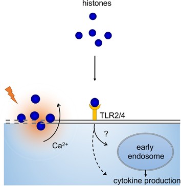
Extracellular histones, cell-free DNA, or nucleosomes: differences in immunostimulation
de
por adulto (o preço varia de acordo com o tamanho do grupo)

